Robert Lombardo spoke at 'Veterans Coffee House' Featured on News 8

Robert Lombardo speaking at ‘Veterans Coffee House’ meet on October 26, 2021.

October 26, 2021- Robert Lombardo, a Vietnam combat veteran with five Purple Heart and two Bronze Stars, spoke about his experience as a Veteran, detailing some close calls he had encountered while in service.

Lombardo is also an adjunct Professor at Quinnipiac University with Diagnostic Imaging.

News 8 captured Lombardo during one of the monthly ‘Veterans Coffee House’ meets hosted at the Wallingford and Cheshire senior centers.

Watch the News 8 clip here.

Summer Research Opportunity for Undergraduates!

We are pleased to announce a new National Science Foundation Research Experiences for Undergraduates (NSF REU) opportunity, sponsored by NSF, the University of South Alabama and Quinnipiac University – The Bioarchaeology of Bronze Age Social Systems. 

Students accepted into the 8-week program will learn to conduct hands-on research with archaeological human skeletal remains from Bronze Age Arabia, receive mentorship from guest scientists, and engage in public outreach.  Student travel (up to $450), housing, and field trip fees will be covered by the NSF, in addition to a $500/week stipend. Eight Fellows will be selected from the pool of applicants. According to NSF eligibility requirements, students must be a US citizen or permanent resident, and currently enrolled in an undergraduate program. 

The Bioarchaeology of Bronze Age Social Systems project will focus on two large Bronze Age skeletal collections from the Umm an-Nar period (2700-2000 BCE) of the United Arab Emirates. An analysis of these skeletons presents an opportunity to examine the socioeconomic, political, and environmental circumstances in which populations in southeastern Arabia resisted stratification, adapted to environmental change, and negotiated their own identities.

For more information about the project and field school check out our website (https://www.southalabama.edu/colleges/artsandsci/syansw/anthropology/reu/research.html)

Website and Online Applicationhttp://goo.gl/kgCi1B

Application Deadline: March 23, 2019

Field School Dates: May 22 – July 15, 2019

Field School Location: University of South Alabama, located in Mobile, AL

Contact Information: Dr. Lesley Gregoricka (lgregoricka@southalabama.edu) and Dr. Jaime Ullinger (jaime.ullinger@qu.edu)

Quinnipiac radiology professors and students use technology to examine Egyptian animal mummies with Egyptologist

NORTH HAVEN — Most things don’t last, unless they’ve been mummified.

Hundreds of 2,000-year-old animal mummies, including crocodiles, cats, baby gazelles, falcons and ibises were removed from storage at Yale’s Peabody Museum of Natural History and brought to Quinnipiac University’s North Haven campus for study by renowned Egyptologist and animal mummy specialist Dr. Salima Ikram, of American University in Cairo and a visiting professor at Yale University.

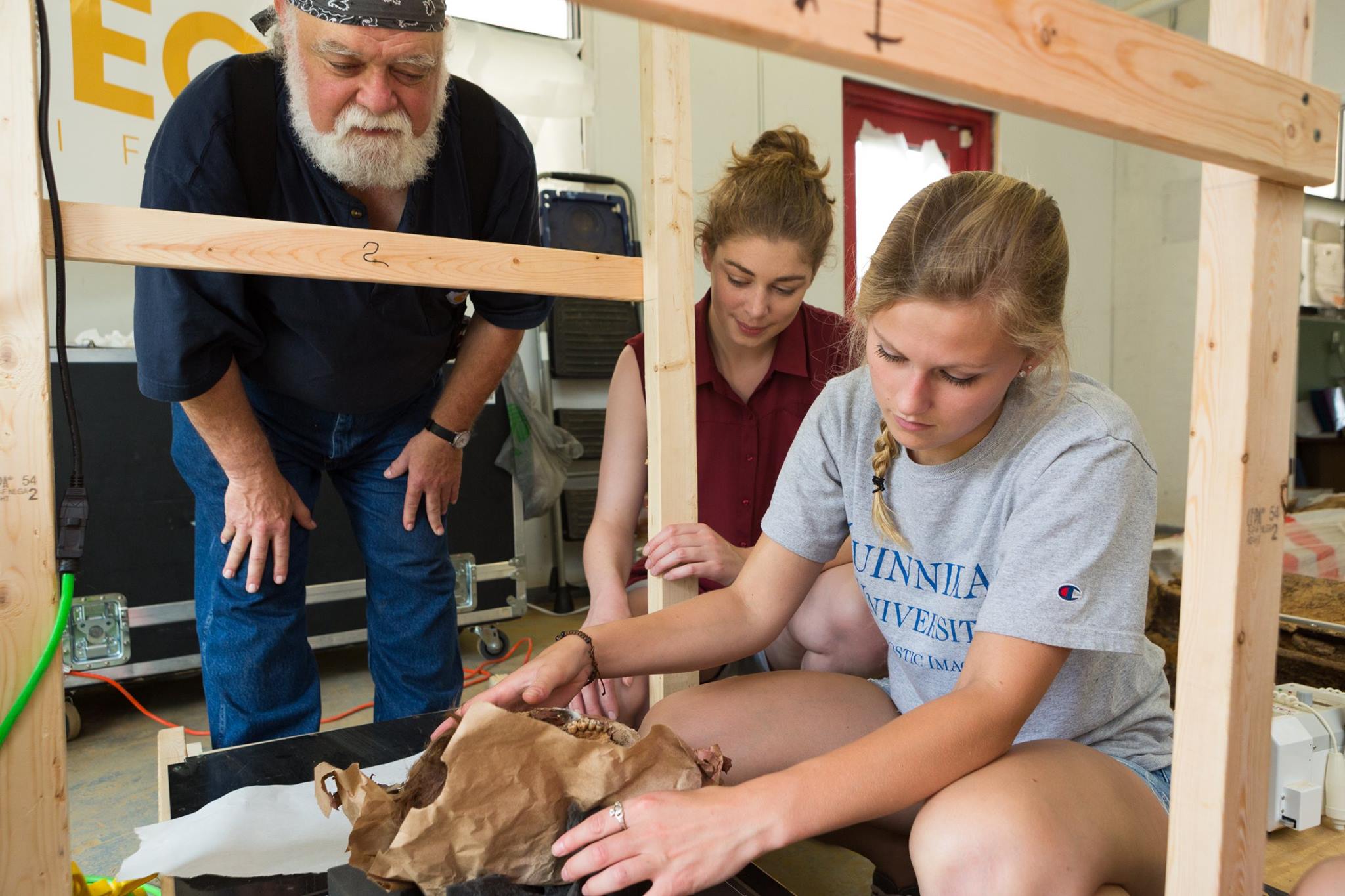
Ikram led Gerald Conlogue, professor emeritus of diagnostic imaging and co-director of the Bioanthropology Research Institute at Quinnipiac University, and William Hennessy, a clinical professor of radiologic science, in the opening of each mummy along with Dr. Roger Colten, collections manager in the division of anthropology for the Peabody.

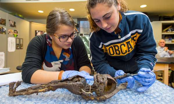
Radiological science, diagnostic imaging and anthropology students from Quinnipiac assisted in in the mummies’ examination using digital radiography, mammography and multi-detector computed tomography. They, along with Near Eastern studies students from Yale, got real-world research experience as the team used the series of machines to inspect each animal mummy, gaining information about the life of the animal before it died and the methods of mummification used.

“This will make a huge difference for us and I also want my students to understand how to unify science and the humanities and to work things out together,” Ikram said. “It’s a good way of unifying the two aspects of ancient Egypt.”

The mummies they are studying would have been wrapped more than 2,000 years ago and used in funeral ceremonies. Mummification of animals in ancient Egypt had several purposes. Sacred animals were bred with the intention of mummification, which are known as votive offerings that were placed in a tomb, but pets were also mummified, Ikram said.

Some animals were mummified to be guardians in the afterlife or at the entrance of a tomb, but they were also mummified as gods because the ancient Egyptians used to believe the spirit of the god would enter into the animal and when it died the spirit of the god would enter another animal, Ikram said.

Hennessy and Conlogue X-rayed these animal mummies 20 years ago with plain film machines, but with the latest digital machines they are able to compare the technological advances and hopefully discover new information.

“Our goal is to take it to the next stage,” Hennessy said. “Is there anything we can see now that we didn’t see 20 years ago because we have better technology? It’s going to provide a lot of information. Time will tell how much information we have, but I think we can do a lot more than we did 20 years ago.”

Film still gives best resolution, he said, but the difference with digital X-rays is a technician can change the levels of contrast of the image so it makes it appear that one can see the image better. With the CT scan a technician can take sectional images, looking at a piece at a time. Notably, the CT scan can look at soft tissue, which wasn’t possible when the mummies were first X-rayed in 1997.

“That’s the magic in the MDCT,” Conlogue said, who also co-hosted the National Geographic program “The Mummy Road Show.” The CT also tells how the animals were wrapped and mummified and shows organs.

“Most excitedly it can tell us its last meal,” Ikram said. In studying the images, Ikram will be looking at mummification methods as well as age and sex and any indication of trauma on the bones because it will help give her a better understanding of the animal’s biography and see how mummification on a large scale in temples was practiced.

“We’re trying to look into that to understand what the ancient Egyptians were thinking and their religious practices in addition to physical stuff,” Ikram said. By studying the new images, she’ll gain information about animal management and animal husbandry in ancient Egypt and be able to better inform a larger picture of the period and people. Ikram has studied similar mummies all over the world since 1996.

“It’s a tremendous opportunity for students to take what they’re learning in the classroom that they have yet to apply, since most have not been in clinic,” Hennessy said.

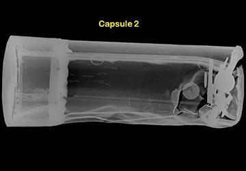
Tania Grgurich, clinical associate professor of diagnostic imaging at Quinnipiac, helped students use the MDCT to image a 2,200-year-old mummified crocodile, creating a 3D image of the mummy. And while that particular crocodile wasn’t wrapped, Grgurich said the machine is able to virtually peel away wrappings on mummies so researchers and technicians can examine the mummy without damaging it.

“It gives people an opportunity to see that as an X-ray technician, you don’t just have to sit here and do live people,” Amanda Taft said, who is a master’s student in health sciences. “You can do mummies and work with anthropologists and things you would never expect.” She assisted in using mammography to examine baby crocodile mummies because it gives a greatly detailed picture.

The images will be sent to Dr. Anthony Fischetti, head of diagnostic imaging and radiology at the Animal Medical Center in Manhattan and a consultant for the Brooklyn Museum’s Soulful Creatures Animal Mummies in Ancient Egypt exhibit. He will determine the age and health of each animal at the time of its death using the new images.

“It’s Halloween and we have mummies walking around,” Hennessy said.

The participating students will assist Conlogue in preparing the technical aspects of the study for presentation along with Ikram’s findings at the World Congress on Mummy Studies in May.

Source: http://www.nhregister.com/metro/article/Quinnipiac-radiology-professors-and-students-use-12321420.php

Colonial Coffins Provide Radiologic Science Students Window to the Past

ColonialCoffinsAug2017.jpg

A team of radiologic sciences students had the opportunity to apply the skills they developed in the classroom for something vastly different than disease diagnosis and treatment last month.

The Quinnipiac students traveled to Burlington, New Jersey with students from the University College Dublin, where they took X-rays of coffins and human remains dating as far back as the early 1700s. The coffins were a major archaeological discovery, and made national headlines when they were excavated from a construction site in the Old City neighborhood of Philadelphia in March.  

“They are more than just boxes of bones,” said Gerald Conlogue, professor emeritus of diagnostic imaging and director of the Bioanthropology Research Institute at Quinnipiac. “They provide us with a picture of both a culture and a time period.”  Conlogue, who led the trip, has been using medical imaging techniques to study mummified and skeletal remains and artifacts for decades. He has worked on archeological sites from Lithuania to Peru, and regularly brings students along on projects.  “I want to broaden their perspectives, and show them that there are many other fields they can take their skills into,” he said.  

Emily Paul ’18, a radiologic sciences major, recalls the excitement of arriving to find “artifacts everywhere,” a full skeleton being recovered by archeologists and state-of-the-art imaging equipment waiting for her.  

Source: https://www.qu.edu/life/now/colonial-coffins-provide-radiologic-sciences-students-window-to-the-past.html

Students Embark on Adventure in United Arab Emirates

QU at Unar 2.jpg

A professor and two Quinnipiac students traveled 7,000 miles during winter break to dive into the “mother of fire.”

While it sounds like the next movie in the Indiana Jones franchise, the excursion to the United Arab Emirates was just as exciting for students Erika Danella '19 and Whitney Wachtarz '18, and Jaime Ullinger, assistant professor of anthropology. The trio spent three weeks sorting and evaluating bones from the tombs of Umm an-Nar, a Bronze Age culture and time period—also known as the “mother of fire”—that existed from 2600-2000 BC in modern-day UAE.

Erika Danella '19, Whitney Wachtarz '18 and Professor Jaime Ullinger is Quinnipiac’s first research group to travel to the United Arab Emirates.

Their team also included Professor Lesley Gregoricka of the University of South Alabama and two of her students and was the first all-female group to conduct research at the site. That fact made local leaders take notice and prompted an invitation to meet with His Highness Sheikh Saud bin Saqr al-Qasimi, Ruler of the Emirate of Ras al-Khaimah and a member of the Federal Supreme Council of the United Arab Emirates.

“He was incredibly welcoming and incredibly supportive of our project,” said Ullinger, who said both the Sheikh and the community are interested in learning more about this time period in their history.

“Drinking coffee with him and eating fresh dates from his royal orchards was a pretty phenomenal experience,” Ullinger said.

The students were impressed by the beautiful palace. Danella, a biology major and anthropology minor, said, “It was cool to talk to him about the work we were doing, and he was really interested.”

Ullinger received Quinnipiac’s Galpin International Exchange Faculty Fellows grant, which helped fund the trip. The students took an independent research class with Ullinger in the fall to prepare.

Using a variety of bioarchaeological techniques, the team endeavored to learn more about the diet, health and behavior of the Umm an-Nar people and their mortuary practices, which included burying large numbers of people in communal tombs.

The team sorted and began to identify a collection of bones that had been excavated in the late 1990s. Most of the bones exist in fragments, Ullinger said, noting that it is possible to identify the age and gender of the person they belonged to.

“The bones can tell us what people were eating, how the environment affected their health, and how they interacted with their environment over time. We also can compare the health of people who had access to water year-round to the bones of people living in other areas who did not,” Ullinger said.

She pointed out that chemical analysis of teeth also can provide clues––from cavities, dental abscesses and absence of teeth lost before death.

Ullinger said the students applied knowledge gleaned in an anatomy class to a real-life situation. “We got through both bone collections, and we accomplished what we needed to. The students worked hard and did a great job.”

Wachtarz, a criminal justice major and anthropology minor, said working onsite was more difficult than she anticipated. “When you are dealing with bones that have been burned, they are warped, and some cannot be identified,” she noted. Sometimes, the students would sort bones into a less relevant pile only to find out later that those bones held important information.

This trip laid the foundation for a long-term collaborative project with researchers in the United Arab Emirates. In addition to new research skills, the students gained a better understanding of the region and its culture by visiting museums and touring other archaeological sites.

“Simply interacting with other people and understanding our commonalities beyond our differences was pretty important,” Ullinger said.

Both professors are collaborating on a National Science Foundation grant that would fund further research opportunities for undergraduate students at the UAE site. Some of those bones will be traveling to Quinnipiac, where the group will work with the university’s Bioanthropology Research Institute, known for providing non-destructive digital imaging to anthropological samples.

“We hope to use X-ray, CT and Micro-CT to understand more about internal structures in bone without having to damage the bone. We can see things like healed fractures and disease processes that would not otherwise be visible,” Ullinger said.

Source: https://www.qu.edu/life/now/research-team-evaluates-bones-in-united-arab-emirates.html

Professor earns $15,000 grant to lead research on Gangi mummies in Italy

Ron Beckett (at right) explains his finding from a past mummy study

Ron Beckett (at right) explains his finding from a past mummy study

Dec. 11, 2014 - A Quinnipiac University professor will lead a team of researchers and students to Italy in January to perform the first-ever examination of the Gangi mummies, whose remains have been preserved in burial catacombs in Sicily.

Ronald Beckett, professor emeritus in biomedical sciences and co-director of the Bioanthropology Research Institute at Quinnipiac, has received a $15,000 grant from the National Geographic Society/Waitt Grants Program to perform paleoimaging and a bioarchaeological analysis of the remains of 60 Gangi mummies from Jan. 1-10.

"While much is known about the origin of these mummies, little is known about their unique wax masks and how they interacted with their environment when they were living and walking the earth," said Beckett, who will serve as the principal investigator on the project. "This expedition will answer a multitude of questions. Who were the Gangi mummies? What diseases did they suffer from? What was their dental condition? Was there evidence of trauma? Why were the wax masks used? How old were they when they died? What modern diseases (osteoporosis, vascular hardening, atherosclerosis, tuberculosis) were present? What is their current state of preservation and the extent of their deterioration?"

In addition to Beckett, the team will include Jerry Conlogue, professor of diagnostic imaging and co-director of the Bioanthropology Research Institute; Dario Piombino-Mascali, scientific curator at the Capuchin Catacombs of Palermo, honorary inspector for the Cultural Heritage of Sicily and director of the "Sicily Mummy Project;" Mark Viner of Cranfield University, the Defence Academy of the United Kingdom, a fellow of the Cranfield Forensic Institute and chief executive officer of the Inforce Foundation; and Katherine Harper-Beckett, a research associate with the Bioanthropology Research Institute. Three diagnostic imaging students at Quinnipiac, Aniello Catapano, Jennifer Curry and Annamaria DiCesare, also will assist the research team.

The NGS/Waitt Grants Program supports cutting-edge research projects in the initial search and exploration phase, when funding is most difficult to secure. Approximately 100 grants of $5,000 to $15,000 will be made annually to explorers and scientists in research fields such as biology, anthropology and the geosciences, who are working across disciplines and responding quickly to potential discoveries.

Created in 1998, the Bioanthropology Research Institute at Quinnipiac conducts research in biology, archaeology, anthropology and paleopathology through paleoimaging applications including diagnostic imaging, video endoscopy, photography and laboratory analysis. The research is conducted on mummified humans and animals and ancient artifacts without destruction. The co-directors collaborate with bioanthropological researchers from around the globe. The institute conducts workshops, gives presentations and conducts field paleoimaging research worldwide.


BMS Center, anthropology program host Hamden students

Senior Alex Burgos shows a group of Hamden Middle School seventh-graders a fossil cast of a skull as part of science and anthropology activities at the University on Oct. 29.

Senior Alex Burgos shows a group of Hamden Middle School seventh-graders a fossil cast of a skull as part of science and anthropology activities at the University on Oct. 29.

Oct. 29, 2013 - Senior Lauren Kaufman is a history major in the five-year education program with an anthropology minor. She got to experience the best of both worlds Oct. 29 as a group of 32 seventh-graders from Hamden Middle School visited the University to learn about science and anthropology.

"I'm getting to combine my two favorite things, which are education and anthropology," said Kaufman, who manned a human osteology station. "It's nice to be able to teach anthropological theories to younger kids so they can grow up thinking that way. It's also exposing them to things that they've never seen in their middle school before. It's exciting to watch them discover something new."

For the second straight year, the Bristol-Myers Squibb Center for Science, Teaching and Learning and the anthropology program teamed up to host the middle-schoolers. The students were divided into two groups and rotated between a pair of classrooms to take part in hand-on activities about archaeological digs, human evolution and technology.

Linda Salters, a seventh-grade teacher at Hamden Middle School, said the science stations certainly held the children's interested.

"They're really enjoying this," Salters said. "They get to be scientists for the day. They ask a lot of questions and the staff at Quinnipiac is very nice. The kids always walk away with something, whether it's a little gift or information they didn't come here with. It's a very nice activity."

The event was organized by Julia Giblin, assistant professor of anthropology, Lucy Howell, director of the Bristol-Myers Squibb Center for Science, Teaching and Learning, and Jaime Ullinger, assistant professor of anthropology and co-director of the Bioanthropology Research Institute

Ullinger said the interactive activities grabbed the children's attention and made learning fun.

"It's super hands on and they're really engaged," she said, adding that the Quinnipiac students get as much out of the day of learning as the middle school students do. "Our students have to use the material they've learned in the classroom and teach it to others. For them, it's a real sense of ownership over the material."

While the Hamden students sifted through sand and examined bones in an anthropology classroom, another group worked with Howell in the Bristol-Myers Squibb Center. She explained technology and had the children build towers out of marshmallows, string, tape and uncooked spaghetti.

"What will future anthropologists and archaeologists think of our culture when they dig up artifacts about our time?" she asked the students to ponder at the end of the activity. 

Diagnostic imaging professor scans 100-year old time capsules

A scan of one of the 100-year old time capsules found when a tree on the New Haven Green was uprooted during the recent hurricane.

A scan of one of the 100-year old time capsules found when a tree on the New Haven Green was uprooted during the recent hurricane.

May 2, 2013 - When New Haven officials discovered a pair of 100-plus-year-old time capsules encased in concrete beneath a Lincoln Oak Tree uprooted during Superstorm Sandy on the New Haven Green, they knew whom to call.

Gerald Conlogue, professor of diagnostic imaging and co-director of the University’s Bioanthropology Research Institute, is renowned for unlocking history’s secrets in some of the most remote corners of the globe.

Conlogue worked with Katelyn D’Alleva and Nini Shingleton, both diagnostic imaging juniors at Quinnipiac, to determine the contents and locations of the 104-year-old 10-inch long by 4-inch wide copper capsules.

“It was imperative to determine the precise locations of each of the 30 items between the two capsules to ensure that they would not be damaged when opening them,” Conlogue said. “Katelyn and Nini had to use all of the knowledge that they developed in the classroom to determine what was inside – and where each item was located. They did a really great job.”

The students helped to take more than 30 X-rays of the capsules to determine the ideal place to enter. They then helped to open the containers.

“It was a very difficult process,” said Drew Days, proprietor of the New Haven Green. “Quinnipiac has been critical in this effort. They came with the sophisticated instruments needed to go into the base and see what was inside.”

Days said without the efforts of Conlogue and his students, “it would have been impossible to bring these up.”

The first capsule was sealed on Feb. 12, 1909 in commemoration of the 100th anniversary of the birth of President Abraham Lincoln. It contained six newspapers, letters and artifacts from the Battle at Gettysburg. The second capsule was sealed on April 9, 1909 in commemoration of the 44th anniversary of General Robert Lee’s surrender at the Battle of Appomattox Court House. It contained commemorative coins, programs and prayer cards, newspapers, a war medal and business cards.

“Quinnipiac was critical in determining where everything was,” said Connecticut State Archaeologist Nicholas Bellantoni.

“It was pretty awesome,” said D’Alleva. “It was a once-in-a-lifetime opportunity to be a part of this.”

Shingleton said the experiences helped her to better appreciate what she has learned – and what she could do with her experience. “It was great to be part of history,” she said. “Professor Conlogue’s passion for this really comes across. He made it a very exciting experience.”

Link for more info:

http://www.pressherald.com/2013/05/05/time-capsules-honor-lincoln-showcase-1909_2013-05-05/