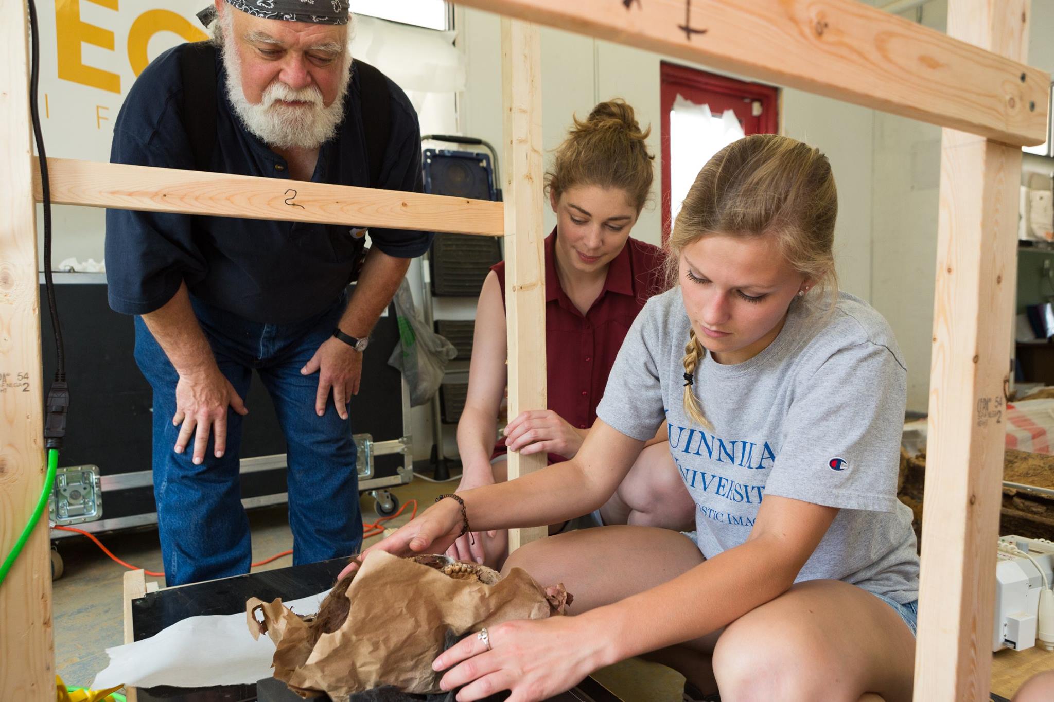
Jaime Ullinger

Robert Lombardo spoke at 'Veterans Coffee House' Featured on News 8

Robert Lombardo speaking at ‘Veterans Coffee House’ meet on October 26, 2021.

October 26, 2021- Robert Lombardo, a Vietnam combat veteran with five Purple Heart and two Bronze Stars, spoke about his experience as a Veteran, detailing some close calls he had encountered while in service.

Lombardo is also an adjunct Professor at Quinnipiac University with Diagnostic Imaging.

News 8 captured Lombardo during one of the monthly ‘Veterans Coffee House’ meets hosted at the Wallingford and Cheshire senior centers.

Watch the News 8 clip here.

Share

Summer Research Opportunity for Undergraduates!

We are pleased to announce a new National Science Foundation Research Experiences for Undergraduates (NSF REU) opportunity, sponsored by NSF, the University of South Alabama and Quinnipiac University – The Bioarchaeology of Bronze Age Social Systems. 

Students accepted into the 8-week program will learn to conduct hands-on research with archaeological human skeletal remains from Bronze Age Arabia, receive mentorship from guest scientists, and engage in public outreach.  Student travel (up to $450), housing, and field trip fees will be covered by the NSF, in addition to a $500/week stipend. Eight Fellows will be selected from the pool of applicants. According to NSF eligibility requirements, students must be a US citizen or permanent resident, and currently enrolled in an undergraduate program. 

The Bioarchaeology of Bronze Age Social Systems project will focus on two large Bronze Age skeletal collections from the Umm an-Nar period (2700-2000 BCE) of the United Arab Emirates. An analysis of these skeletons presents an opportunity to examine the socioeconomic, political, and environmental circumstances in which populations in southeastern Arabia resisted stratification, adapted to environmental change, and negotiated their own identities.

For more information about the project and field school check out our website (https://www.southalabama.edu/colleges/artsandsci/syansw/anthropology/reu/research.html)

Website and Online Applicationhttp://goo.gl/kgCi1B

Application Deadline: March 23, 2019

Field School Dates: May 22 – July 15, 2019

Field School Location: University of South Alabama, located in Mobile, AL

Contact Information: Dr. Lesley Gregoricka (lgregoricka@southalabama.edu) and Dr. Jaime Ullinger (jaime.ullinger@qu.edu)

Share

Quinnipiac radiology professors and students use technology to examine Egyptian animal mummies with Egyptologist

NORTH HAVEN — Most things don’t last, unless they’ve been mummified.

Hundreds of 2,000-year-old animal mummies, including crocodiles, cats, baby gazelles, falcons and ibises were removed from storage at Yale’s Peabody Museum of Natural History and brought to Quinnipiac University’s North Haven campus for study by renowned Egyptologist and animal mummy specialist Dr. Salima Ikram, of American University in Cairo and a visiting professor at Yale University.

Ikram led Gerald Conlogue, professor emeritus of diagnostic imaging and co-director of the Bioanthropology Research Institute at Quinnipiac University, and William Hennessy, a clinical professor of radiologic science, in the opening of each mummy along with Dr. Roger Colten, collections manager in the division of anthropology for the Peabody.

Radiological science, diagnostic imaging and anthropology students from Quinnipiac assisted in in the mummies’ examination using digital radiography, mammography and multi-detector computed tomography. They, along with Near Eastern studies students from Yale, got real-world research experience as the team used the series of machines to inspect each animal mummy, gaining information about the life of the animal before it died and the methods of mummification used.

“This will make a huge difference for us and I also want my students to understand how to unify science and the humanities and to work things out together,” Ikram said. “It’s a good way of unifying the two aspects of ancient Egypt.”

The mummies they are studying would have been wrapped more than 2,000 years ago and used in funeral ceremonies. Mummification of animals in ancient Egypt had several purposes. Sacred animals were bred with the intention of mummification, which are known as votive offerings that were placed in a tomb, but pets were also mummified, Ikram said.

Some animals were mummified to be guardians in the afterlife or at the entrance of a tomb, but they were also mummified as gods because the ancient Egyptians used to believe the spirit of the god would enter into the animal and when it died the spirit of the god would enter another animal, Ikram said.

Hennessy and Conlogue X-rayed these animal mummies 20 years ago with plain film machines, but with the latest digital machines they are able to compare the technological advances and hopefully discover new information.

“Our goal is to take it to the next stage,” Hennessy said. “Is there anything we can see now that we didn’t see 20 years ago because we have better technology? It’s going to provide a lot of information. Time will tell how much information we have, but I think we can do a lot more than we did 20 years ago.”

Film still gives best resolution, he said, but the difference with digital X-rays is a technician can change the levels of contrast of the image so it makes it appear that one can see the image better. With the CT scan a technician can take sectional images, looking at a piece at a time. Notably, the CT scan can look at soft tissue, which wasn’t possible when the mummies were first X-rayed in 1997.

“That’s the magic in the MDCT,” Conlogue said, who also co-hosted the National Geographic program “The Mummy Road Show.” The CT also tells how the animals were wrapped and mummified and shows organs.

“Most excitedly it can tell us its last meal,” Ikram said. In studying the images, Ikram will be looking at mummification methods as well as age and sex and any indication of trauma on the bones because it will help give her a better understanding of the animal’s biography and see how mummification on a large scale in temples was practiced.

“We’re trying to look into that to understand what the ancient Egyptians were thinking and their religious practices in addition to physical stuff,” Ikram said. By studying the new images, she’ll gain information about animal management and animal husbandry in ancient Egypt and be able to better inform a larger picture of the period and people. Ikram has studied similar mummies all over the world since 1996.

“It’s a tremendous opportunity for students to take what they’re learning in the classroom that they have yet to apply, since most have not been in clinic,” Hennessy said.

Tania Grgurich, clinical associate professor of diagnostic imaging at Quinnipiac, helped students use the MDCT to image a 2,200-year-old mummified crocodile, creating a 3D image of the mummy. And while that particular crocodile wasn’t wrapped, Grgurich said the machine is able to virtually peel away wrappings on mummies so researchers and technicians can examine the mummy without damaging it.

“It gives people an opportunity to see that as an X-ray technician, you don’t just have to sit here and do live people,” Amanda Taft said, who is a master’s student in health sciences. “You can do mummies and work with anthropologists and things you would never expect.” She assisted in using mammography to examine baby crocodile mummies because it gives a greatly detailed picture.

The images will be sent to Dr. Anthony Fischetti, head of diagnostic imaging and radiology at the Animal Medical Center in Manhattan and a consultant for the Brooklyn Museum’s Soulful Creatures Animal Mummies in Ancient Egypt exhibit. He will determine the age and health of each animal at the time of its death using the new images.

“It’s Halloween and we have mummies walking around,” Hennessy said.

The participating students will assist Conlogue in preparing the technical aspects of the study for presentation along with Ikram’s findings at the World Congress on Mummy Studies in May.

Source: http://www.nhregister.com/metro/article/Quinnipiac-radiology-professors-and-students-use-12321420.php
Share

Colonial Coffins Provide Radiologic Science Students Window to the Past

ColonialCoffinsAug2017.jpg

A team of radiologic sciences students had the opportunity to apply the skills they developed in the classroom for something vastly different than disease diagnosis and treatment last month.

The Quinnipiac students traveled to Burlington, New Jersey with students from the University College Dublin, where they took X-rays of coffins and human remains dating as far back as the early 1700s. The coffins were a major archaeological discovery, and made national headlines when they were excavated from a construction site in the Old City neighborhood of Philadelphia in March.  

“They are more than just boxes of bones,” said Gerald Conlogue, professor emeritus of diagnostic imaging and director of the Bioanthropology Research Institute at Quinnipiac. “They provide us with a picture of both a culture and a time period.”  Conlogue, who led the trip, has been using medical imaging techniques to study mummified and skeletal remains and artifacts for decades. He has worked on archeological sites from Lithuania to Peru, and regularly brings students along on projects.  “I want to broaden their perspectives, and show them that there are many other fields they can take their skills into,” he said.  

Emily Paul ’18, a radiologic sciences major, recalls the excitement of arriving to find “artifacts everywhere,” a full skeleton being recovered by archeologists and state-of-the-art imaging equipment waiting for her.  

Source: https://www.qu.edu/life/now/colonial-coffins-provide-radiologic-sciences-students-window-to-the-past.html

Share

Students Embark on Adventure in United Arab Emirates

QU at Unar 2.jpg

A professor and two Quinnipiac students traveled 7,000 miles during winter break to dive into the “mother of fire.”

While it sounds like the next movie in the Indiana Jones franchise, the excursion to the United Arab Emirates was just as exciting for students Erika Danella '19 and Whitney Wachtarz '18, and Jaime Ullinger, assistant professor of anthropology. The trio spent three weeks sorting and evaluating bones from the tombs of Umm an-Nar, a Bronze Age culture and time period—also known as the “mother of fire”—that existed from 2600-2000 BC in modern-day UAE.

Erika Danella '19, Whitney Wachtarz '18 and Professor Jaime Ullinger is Quinnipiac’s first research group to travel to the United Arab Emirates.

Their team also included Professor Lesley Gregoricka of the University of South Alabama and two of her students and was the first all-female group to conduct research at the site. That fact made local leaders take notice and prompted an invitation to meet with His Highness Sheikh Saud bin Saqr al-Qasimi, Ruler of the Emirate of Ras al-Khaimah and a member of the Federal Supreme Council of the United Arab Emirates.

“He was incredibly welcoming and incredibly supportive of our project,” said Ullinger, who said both the Sheikh and the community are interested in learning more about this time period in their history.

“Drinking coffee with him and eating fresh dates from his royal orchards was a pretty phenomenal experience,” Ullinger said.

The students were impressed by the beautiful palace. Danella, a biology major and anthropology minor, said, “It was cool to talk to him about the work we were doing, and he was really interested.”

Ullinger received Quinnipiac’s Galpin International Exchange Faculty Fellows grant, which helped fund the trip. The students took an independent research class with Ullinger in the fall to prepare.

Using a variety of bioarchaeological techniques, the team endeavored to learn more about the diet, health and behavior of the Umm an-Nar people and their mortuary practices, which included burying large numbers of people in communal tombs.

The team sorted and began to identify a collection of bones that had been excavated in the late 1990s. Most of the bones exist in fragments, Ullinger said, noting that it is possible to identify the age and gender of the person they belonged to.

“The bones can tell us what people were eating, how the environment affected their health, and how they interacted with their environment over time. We also can compare the health of people who had access to water year-round to the bones of people living in other areas who did not,” Ullinger said.

She pointed out that chemical analysis of teeth also can provide clues––from cavities, dental abscesses and absence of teeth lost before death.

Ullinger said the students applied knowledge gleaned in an anatomy class to a real-life situation. “We got through both bone collections, and we accomplished what we needed to. The students worked hard and did a great job.”

Wachtarz, a criminal justice major and anthropology minor, said working onsite was more difficult than she anticipated. “When you are dealing with bones that have been burned, they are warped, and some cannot be identified,” she noted. Sometimes, the students would sort bones into a less relevant pile only to find out later that those bones held important information.

This trip laid the foundation for a long-term collaborative project with researchers in the United Arab Emirates. In addition to new research skills, the students gained a better understanding of the region and its culture by visiting museums and touring other archaeological sites.

“Simply interacting with other people and understanding our commonalities beyond our differences was pretty important,” Ullinger said.

Both professors are collaborating on a National Science Foundation grant that would fund further research opportunities for undergraduate students at the UAE site. Some of those bones will be traveling to Quinnipiac, where the group will work with the university’s Bioanthropology Research Institute, known for providing non-destructive digital imaging to anthropological samples.

“We hope to use X-ray, CT and Micro-CT to understand more about internal structures in bone without having to damage the bone. We can see things like healed fractures and disease processes that would not otherwise be visible,” Ullinger said.

Source: https://www.qu.edu/life/now/research-team-evaluates-bones-in-united-arab-emirates.html

Share